an358-hibit-blotting
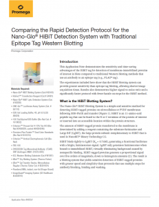
Comparing the Rapid Detection Protocol for the Nano-Glo® HiBiT Detection System with Traditional Epitope Tag Western Blotting Sidebar Headers style: Sidebar Body style Sidebar Headers style: • Sidebar Body style w/bullet (first) • Sidebar Bullets (not first) style Application Note #AN358 Promega Corporation Materials Required: • Nano-Glo® HiBiT Blotting System (Cat.# N2410) • ViaFect™ Transfection Reagent (Cat.# E4981) • Nano-Glo® HiBiT Lytic Detection System (Cat. # N3030) • ONE-Glo™ Luciferase Assay System (Cat. # E6110) • SDS Gel Loading Buffer, 4X, pH 6.8 (LSS AB424) • 10X Tris/Glycine/SDS Buffer (Bio-Rad Cat. # 1610832) • Criterion™ Precast Gel 4-15% Tris-HCl (BioRad #3450029, control #64304246) • Precision Plus Protein™ Dual Color Standards (Bio-Rad #1610374) • Criterion™ Vertical Electrophoresis Cell (BioRad) • TBS 10X • DYKDDDDK Tag Monoclonal Antibody (FG4R) HRP (Invitrogen #MA1-91878-HRP,) • ECL Western Blotting Substrate (Cat.# W1001) • iBlot™ Dry Blotting System (Thermo Fisher) • iBlot™ Gel Transfer Stacks, Nitrocellulose Regular (Thermo Fisher Cat. # IB301001) • Powdered Milk, instant, non-fat (Kroger Brand) • ImageQuant® Imaging System (GE Healthcare #LAS4010) Introduction This Application Note demonstrates the sensitivity and time-saving advantages of the HiBiT tag for detection of membrane-immobilized proteins of interest in blots compared to traditional Western blotting methods that use an antibody to an epitope tag (e.g., FLAG® tag). The experiments included here show that the HiBiT blotting system can provide greater sensitivity than epitope tag blotting, allowing shorter image acquisition times. Results also demonstrate higher signal-to-noise ratio and a significantly faster protocol with fewer hands-on steps for the HiBiT method. What is the HiBiT Blotting System? The Nano-Glo® HiBiT Blotting System is a simple and sensitive method for detecting HiBiT-tagged proteins on nitrocellulose or PVDF membranes following SDS-PAGE and transfer (Figure 1). HiBiT is an 11-amino-acid peptide tag that can be fused to the N or C terminus of the protein of interest or inserted into an accessible location within the protein structure. The amount of HiBiT-tagged protein transferred to the membrane is determined by adding a reagent containing the substrate furimazine and Large BiT (LgBiT), the large protein subunit complementary to HiBiT that is used in NanoBiT® Binary Technology (1). HiBiT binds tightly to LgBiT (KD = 0.7nM), generating a functional luciferase with a bright, luminescence signal. LgBiT only generates luminescence when bound to immobilized HiBiT, virtually eliminating background caused by nonspecific binding. HiBiT-tagged proteins generate a proportional signal over five orders of magnitude, down to femtogram amounts (2). The result is a blotting system that yields sensitive detection of HiBiT-tagged proteins with greater speed and simplicity than protocols that use multiple steps for antibody blocking, binding and washing. 2 Comparing Nano-Glo® HiBiT Blotting with a Traditional Epitope Tag Western Blot Method Promega Corporation Experimental Strategy HEK293 cells were transiently transfected with a plasmid expressing firefly luciferase with C-terminal FLAG® and HiBiT tags (pRSG55). A serial dilution of cell lysates was prepared and separated by electrophoresis on an SDS-PAGE gel. The proteins were transferred to a nitrocellulose membrane, and the blot was split into two halves—one for blotting with the Nano-Glo® HiBiT Blotting System and another for traditional blotting with a horseradish peroxidase-conjugated anti-FLAG® antibody, which was detected by luminescence using the ECL Western Blotting Substrate. Images were generated every minute over 60 minutes using a CCD imager, the ImageQuant® System. Methods Reverse transfection of pRSG55 plasmid into HEK293 cells. Cells were trypsinized and then resuspended in media. Cells were pelleted by centrifugation at 300 × g for 10 minutes, the media was removed, and the cell pellet gently resuspended in media (DMEM + 10% FBS + Pen/Strep). Cells were counted and diluted to 100,000 cells/ml. Transfection complexes were prepared with ViaFect™ Transfection Reagent, using a 6:1 ratio of reagent to DNA (Table 1). Transfection complexes were incubated for 10 minutes, and then brought up to a volume of 100µl with OptiMEM® Reduced Serum Medium. Table 1. Preparation of Transfection Complexes Component Volume (1X) Final Concentration Volume (15X) DNA 1.87µl 1µg 28.0µl OptiMEM Media 8.13µl — 122µl ViaFect 6µl 6:1 90µl Incubate above at room temperature for 10 minutes prior to bringing up to 100µl volume. OptiMEM Media 84µl — 1.26ml 200µl of the pRSG55 transfection complex was added to 5 wells of a 6-well plate. 2ml of cells at 100,000 cells/ml was pipetted on top of the transfection complexes nnd the plate was mixed on a plate shaker at 750rpm for 1 minute. The plate was then placed in a 37°C incubator with 5% CO2 for about 24 hours. 14368MB Separate HiBiT-tagged proteins in cell lysate using SDS-PAGE. Transfer proteins from gel to nitrocellulose membrane. Add Nano-Glo® HiBiT Blotting System reagents to the membrane to detect HiBiT-tagged proteins. Image the membrane. Gel Membrane Figure 1. The Nano-Glo® HiBiT Blotting System method. Samples including a HiBiT-tagged protein are separated by SDS-PAGE and transferred to nitrocellulose or PVDF membranes. LgBiT protein in the reagent binds to the immobilized HiBiT tag and generates a luminescent enzyme in the presence of furimizine. Application Note #AN358 3 Blotting of the dual-tagged HiBiT and FLAG®-tagged luciferase construct. After confirming that the cells had been successfully transfected by measuring luciferase activity (data not shown) 1ml aliquots of pRSG55 transfected cells or 500µl aliquots of control transfection cells were centrifuged at 500 × g for 10 minutes to pellet the cells, the media was removed, and the cell pellets were frozen at –80°C. A thawed pRSG55-transfected cell pellet (100,000 cells) was resuspended in 100µl of 1X SDS loading buffer containing 5% 1-thioglycerol. To lyse cells and denature the proteins, the cell pellet was incubated at 95°C for 5 minutes. The lysate was then serially diluted 1:4 down to the equivalent of about 40 cells/10µl. The control pellet of 100,000 untransfected HEK293 cells was resuspended in 100µl 1X SDS Loading Buffer with 5% 1-thioglycerol and incubated at 95°C for 5 minutes. 10µl of each lysate or lysate dilution was loaded on a 4-15% Tris-HCl precast gel. The gel was run with 1X Tris/Glycine/SDS Buffer at 200V for 1 hour until the bromophenol dye front just ran off the bottom. The proteins were transferred from the gel to a nitrocellulose membrane using the iBlot™ Transfer System with the corresponding nitrocellulose gel transfer stacks. Proteins were transferred using program P3 on the iBlot™ Transfer System with a 7-minute transfer time. The blot was cut in half, and both halves were transferred to separate containers with TBST. HiBiT Blotting For Nano-Glo® HiBiT blotting the Rapid Detection Protocol for Nitrocellulose Membranes was performed as described in the Nano-Glo® HiBiT Blotting System Technical Manual, TM524. The blot was incubated in TBST at room temperature with shaking for 30 minutes to allow for solubilization of the tag. 10ml of Nano-Glo® HiBiT Blotting Reagent (with both LgBiT and the NanoGlo® substrate) was prepared according to the Technical Manual. The blot was incubated with the Nano-Glo® HiBiT Blotting Reagent for 5 minutes, and then imaged using the ImageQuant® System for 60 minutes, with images generated every minute. Epitope Tag Blotting For traditional blotting with an anti-FLAG® antibody conjugated to HRP, the blot was first blocked in TBST + 5% powdered milk solution for 1 hour with shaking at room temperature. The TBST/milk solution was decanted, and the blot was probed with 10 ml of a solution containing a 1:500 dilution (manufacturer’s recommended dilution) of the anti-FLAG® antibody prepared in TBST + 5% milk. The blot was incubated with the antibody solution for 1 hour at room temperature with shaking. Following incubation with the antibody solution, the membrane was rinsed with TBST. The membrane was washed 3X with TBST for a total of 15 minutes of washing. The blot was then incubated with 15ml of the prepared ECL Western Blotting Substrate for 1 minute at room temperature and the blot was imaged using the ImageQuant® System for 60 minutes, with images generated every minute. Results Blotting with the rapid detection protocol using the Nano-Glo® HiBiT Blotting System was much faster, saving an average of approximately 1.5 hours compared to probing with a traditional epitope tag. Images of blots detected with either Nano-Glo® HiBiT Blotting System or the traditional epitope tag blotting with an anti-FLAG® antibody are shown for 1, 5, 15, 30, 45, and 60-minute time points in Figure 2. At the earliest time point, bands corresponding to the luciferase construct are visible for the blot probed with the NanoGlo® HiBiT Blotting System, but not for the blot probed with the anti-FLAG® antibody solution. By 15 minutes, bands corresponding to the lowest dilution of cells (40 cells/lane) were apparent with the HiBiT Blotting System, but not for the anti-FLAG® blot. 4 Comparing Nano-Glo® HiBiT Blotting with a Traditional Epitope Tag Western Blot Method Promega Corporation α-FLAG, HRP antibody Exposure: 1 minute HiBiT Blotting α-FLAG, HRP antibody HiBiT Blotting Exposure: 5 minutes α-FLAG, HRP antibody HiBiT Blotting α-FLAG, HRP antibody HiBiT Blotting α-FLAG, HRP antibody HiBiT Blotting α-FLAG, HRP antibody HiBiT Blotting Exposure: 15 minutes Exposure: 30 minutes Exposure: 45 minutes Exposure: 60 minutes 17155T A Figure 2. Images obtained with the Image Quant System for blots probed with the rapid detection Nano-Glo® HiBiT blotting protocol versus a traditional Western blotting protocol for detection of the FLAG® tag with an anti-FLAG® antibody conjugated to HRP. Cells were transfected with a construct that encodes for a firefly luciferase construct which is dual tagged with both the FLAG® tag sequence and HiBiT sequence on the C-terminus. Images are shown for 1, 5, 15, 30, 45, and 60-minute exposure times. Loaded 10µl of lysate containing the equivalent of 10,000 transfected cells, 2500 cells, 625 cells, 156 cells, and 40 cells as well as a non-transfected control (in that order) duplicated across each blot. To allow for direct comparison of the blots, images were collected with the same settings on the ImageQuant® System, and there is no further image processing such as contrast enhancement. PROMEGA CORPORATION • 2800 WOODS HOLLOW ROAD • MADISON, WI 53711-5399 USA • TELEPHONE 608-274-4330 www.promega.com • ©2020 PROMEGA CORPORATION • ALL RIGHTS RESERVED • PRICES AND SPECIFICATIONS SUBJECT TO CHANGE WITHOUT PRIOR NOTICE • PRINTED IN USA 06/20 • 58675419 PART #AN358 At all time points, the anti-FLAG® blot appears to have a lower signal-to-background ratio than the HiBiT blot because there is less contrast between the bands and the background. The higher background signal is particularly apparent at longer exposure times. For the Nano-Glo® HiBiT Blotting System the intensity of the bands increases with longer exposures, but the blot background does not increase over time. For the anti-FLAG® blot, the background does increase (appears darker in these color inverted images) over time. In contrast, if the LgBiT “probe” adheres to the membrane nonspecifically, it cannot contribute to background signal because the complementation interaction required for enzymatic activity is not fulfilled. This creates cleaner images with better signal-to-background ratios. Additional sensitivity gains may be observed with the slightly longer (by approximately 30 minutes) Standard Detection Protocol for the Nano-Glo® HiBiT Blotting System. However, it was not within the scope of this work to compare the two detection protocols. Conclusions The Nano-Glo® HiBiT Blotting System provides an alternative to traditional epitope-tag blotting methods, allowing for a faster blotting protocol with less hands-on time, while still providing a sensitive method for detection of tagged proteins. In this experiment, the increased sensitivity observed with the Nano-Glo® HiBiT Blotting System allowed for shorter image acquisition times, further decreasing the time to results. The complementation-based detection method used in HiBiT blotting eliminates the need to block and wash the membrane and results in very little background or nonspecific bands, offering improved signal-to-noise ratios and very clean blotting results. References 1. Dixon, A.S. et al. (2016) NanoLuc complementation reporter optimized for accurate measurement of protein interactions in cells. ACS Chem. Biol. 11, 400–8. 2. Nano-Glo® HiBiT Blotting System Technical Manual, TM524, Promega Corporation. Ordering Information Product Size Cat.# Nano-Glo® HiBiT Blotting System 100ml N2410 ViaFect is a trademark of Promega Corporation. One-Glo and Nano-Glo are registered trademarks of Promega Corporation. Criterion is a trademark of Bio-Rad Corporation. FLAG is a trademark of Sigma-Aldrich Biotechnology LP and ANTI-FLAG is a trademark of Sigma-Aldrich Co.LLC. iBlot and OptiMEM are trademarks of Thermo Fisher Scientific. ImageQuant is a trademark of GE Healthcare Bio Sciences. Products may be covered by pending or issued patents or may have certain limitations. Please visit www.promega.com for details.